Explore
Featured
Recent
Articles
Topics
Login
Upload
Featured
Recent
Articles
Topics
Login
Upload
Search Results for 'cell figure'
cell figure published presentations and documents on DocSlides.
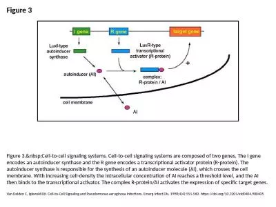
Figure 3 Figure 3. Cell-to-cell signaling systems. Cell-to-cell signaling systems are comp
by pagi
Van Delden C, Iglewski BH. Cell-to-Cell Signaling ...
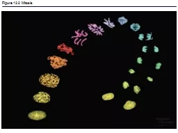
Figure 12.0 Mitosis Figure 12.1a The functions of cell division: Reproduction
by sistertive
Figure 12.1b The functions of cell division: Grow...
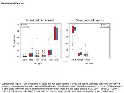
Supplemental Figure 4 Supplemental Figure 4: Cell proportions for septic and non-septic patients i
by lucinda
drived. using the Houseman deconvolution method [...
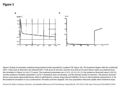
Figure 5 Figure 5. 5a simulates combined drug treatment data reported for a patient (18; F
by ximena
Kirschner DE, Webb G. Resistance, Remission, and Q...
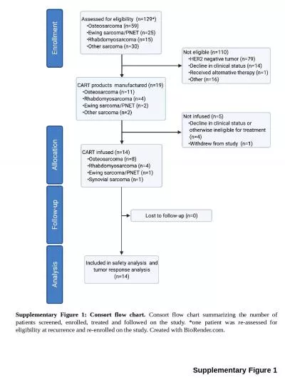
Supplementary Figure 1 Supplementary Figure 1: Consort flow chart.
by leah
Consort flow chart summarizing the number of patie...
Figure 23.1
by danika-pritchard
Figure 23.3. Human Microbiota – Respiratory Tra...
Figure 12.0 Mitosis
by alexa-scheidler
Figure 12.1a The functions of cell division: Rep...
Lecture 14 Cell Communication
by evelyn
Outline. Review of Photosynthesis. Finish up Cam p...
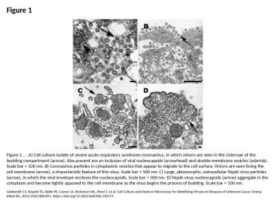
Figure 1 Figure 1. . . A) Cell culture isolate of severe acute respiratory syndrome coronavirus, in
by davies
Goldsmith CS, Ksiazek TG, Rollin PE, Comer JA, Nic...
Figure S1 . Effect of avutometinib on cell viability of NCI-H358 cells.
by amelia
The NCI-H358 cells were treated with avutometinib ...
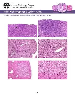
Basophilic Eosinophilic Clear cell Mixed Focus
by cadie
Liver Figure Legend:Figure Basophilic focus (arrow...
Concepts of the Cell Theory
by test
A cell is the basic structural and functional uni...
Figure 25.0 -1 Why Hormones
by fanny
Matter. Figure . 25.0. -. 1a. Figure . 25.0. -. 1b...
Figure 18.2 Regulation of gene
by fluental
expression. Feedback. inhibition. Precursor. Gene...
Figure 17-5b The Sectional Anatomy of the Eye.
by olivia-moreira
Posterior. cavity. Iris. Ciliary. body. Choroid....
Review Unit 3 and 4 Figure 6.8a
by olivia-moreira
ENDOPLASMIC RETICULUM (ER). Rough. ER. Smooth. ER...
Figure 26.0-1 Why Reproduction Matters
by phoebe-click
Figure 26.0-1a. Figure 26.0-1b. Figure 26.0-1ba. ...
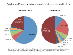
Supplemental Figure 1. Mutation frequencies in adenocarcinoma of the lung
by giovanna-bartolotta
Data source: . Wang R, Pan Y, Li C, et al. Analys...
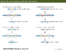
Figure 2.26 Some examples of alternative RNA splicing
by test
Figure 2.31 Degradation of casein mRNA in the pr...
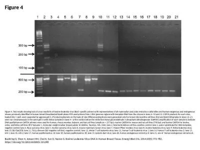
Figure 4 Figure 4. Test results showing lack of cross-reactivity of bovine leukemia virus (BLV)–s
by titus331
Buehring G, Shen H, Jensen HM, Choi K, Sun D, Nuov...
Figure 2 Figure 2. . . A) Extracellular lymphocytic choriomeningitis virus particles (arrow) contai
by carla
Goldsmith CS, Ksiazek TG, Rollin PE, Comer JA, Nic...
Zoo-352 Principles of genetics
by isla
Lecture 4. Mitosis. Mitosis is a part of the . cel...
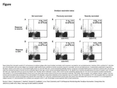
Figure Figure. Flow cytometric analysis of T-cell responses to smallpox antigens after rec
by patricia
Poccia F, Gioia C, Montesano C, Martini F, Horejsh...
Chapter 15: HIV and AIDS
by jalin
Biology Trending, 4e. Eli Minkoff and Jennifer Hoo...
Prepare for chapter three – watch this tutorial before class weds the 17th
by badra
https://sophia.smith.edu/blog/barresilab/devidetor...
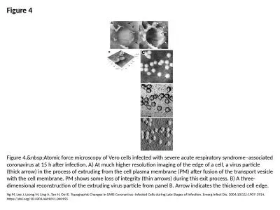
Figure 4 Figure 4. Atomic force microscopy of Vero cells infected with severe acute respir
by ella
Ng M, Lee J, Leong M, Ling A, Tan H, Ooi E. Topogr...
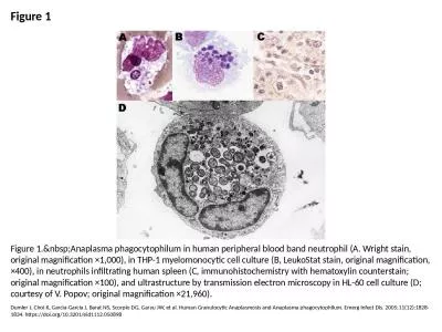
Figure 1 Figure 1. Anaplasma phagocytophilum in human peripheral blood band neutrophil (A.
by skylar
Dumler J, Choi K, Garcia-Garcia J, Barat NS, Scorp...
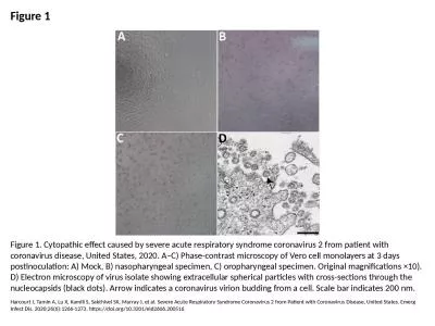
Figure 1 Figure 1. Cytopathic effect caused by severe acute respiratory syndrome coronavirus 2 from
by lam
Harcourt J, Tamin A, Lu X, Kamili S, Sakthivel SK,...
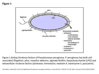
Figure 1 Figure 1. Virulence factors of Pseudomonas aeruginosa. P. aeruginosa has both cel
by lydia
Van Delden C, Iglewski BH. Cell-to-Cell Signaling ...
Unit 5: Subcellular Genetic Elements
by esther
Chapter 23 - Plasmids. Figure 23.01. . Plasmids. ...
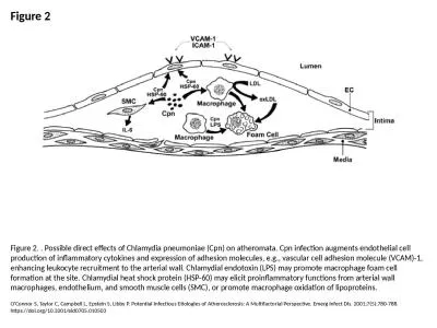
Figure 2 Figure 2. . Possible direct effects of Chlamydia pneumoniae (Cpn) on atheromata. Cpn infec
by cora
O'Connor S, Taylor C, Campbell L, Epstein S, Libby...
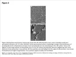
Figure 2 Figure 2. Scanning electron microscopy of Vero E6 cells infected with severe acut
by clara
Ng M, Lee J, Leong M, Ling A, Tan H, Ooi E. Topogr...
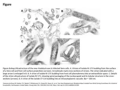
Figure Figure. Ultrastructure of the new rhabdoviruses in infected Vero cells. A. Virions
by morton
Travassos da Rosa AP, Mather TN, Takeda T, Whiteho...
Classification of Eukaryotes
by melanie
5.1 The History of Eukaryotes First eukaryoti...
Unit 6: Changing the DNA Blueprint
by Goofball
Chapter 28 - Bacterial Genetics. Figure 28.01. Rec...
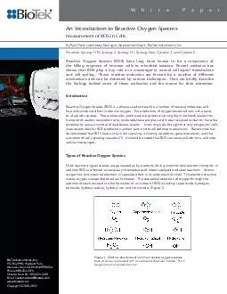
An Introduction to Reactive Oxygen SpeciesMeasurement of ROS in Cells
by grace3
1 White PaperBioTek Instruments IncPO Box 998 High...
Supplementary
by roxanne
Information Discovery of Novel [FeFe] - Hydrogenas...
Progression in Neoplastic Development
by cheryl-pisano
Folder Title: . Progress(. NoTP. ). Updated: Marc...
0 Chapter 9 Mitosis You Must Know
by trish-goza
The features of mitosis that result in the produc...
Gram-Negative Outer Membrane
by stefany-barnette
Figure 4.13c. Figure 4.13b–c. Gram-Positive Gra...
Load More...