Explore
Featured
Recent
Articles
Topics
Login
Upload
Featured
Recent
Articles
Topics
Login
Upload
Search Results for 'figure cells'
figure cells published presentations and documents on DocSlides.
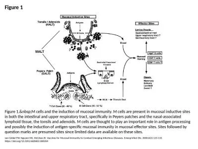
Figure 1 Figure 1. M cells and the induction of mucosal immunity. M cells are present in m
by naomi
van Ginkel FW, Nguyen HH, McGhee JR. Vaccines for ...
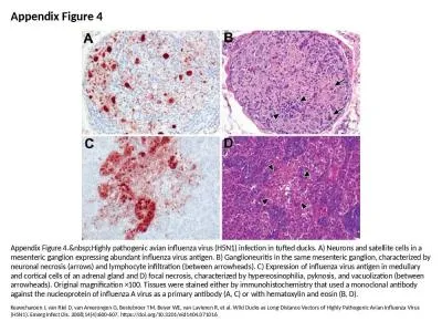
Appendix Figure 4 Appendix Figure 4. Highly pathogenic avian influenza virus (H5N1) infect
by elizabeth
Keawcharoen J, van Riel D, van Amerongen G, Besteb...
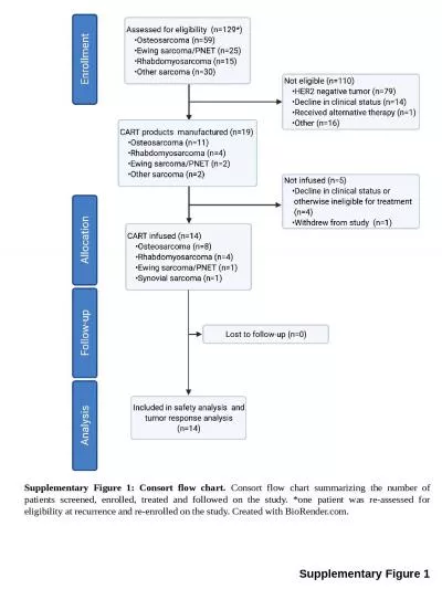
Supplementary Figure 1 Supplementary Figure 1: Consort flow chart.
by leah
Consort flow chart summarizing the number of patie...
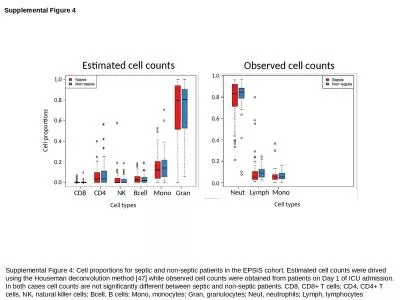
Supplemental Figure 4 Supplemental Figure 4: Cell proportions for septic and non-septic patients i
by lucinda
drived. using the Houseman deconvolution method [...
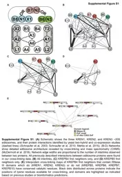
Supplemental Figure S1 Supplemental
by aquaticle
Figure . S1. . . (A) . Schematic shows the three K...
Figure 23.1
by danika-pritchard
Figure 23.3. Human Microbiota – Respiratory Tra...
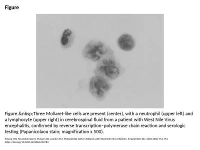
Figure Figure. Three Mollaret-like cells are present (center), with a neutrophil (upper le
by white
Procop GW, Yen-Lieberman B, Prayson RA, Gordon SM....
Figure S1 . Effect of avutometinib on cell viability of NCI-H358 cells.
by amelia
The NCI-H358 cells were treated with avutometinib ...
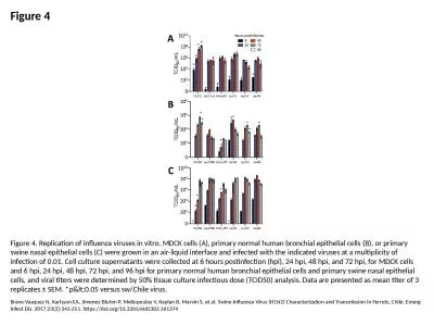
Figure 4 Figure 4. Replication of influenza viruses in vitro. MDCK cells (A), primary normal human
by jasmine
Bravo-Vasquez N, Karlsson EA, Jimenez-Bluhm P, Mel...
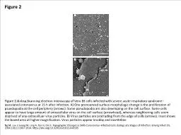
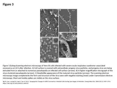
Figure 2 Figure 2. Scanning electron microscopy of Vero E6 cells infected with severe acut
by clara
Ng M, Lee J, Leong M, Ling A, Tan H, Ooi E. Topogr...
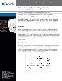
An Introduction to Reactive Oxygen SpeciesMeasurement of ROS in Cells
by grace3
1 White PaperBioTek Instruments IncPO Box 998 High...
Myeloid-Derived Suppressor Cells in Autoimmune Disease
by jane-oiler
By: Chrisanthi Kanaris. Dr. . Lynes. . MCB 5255....
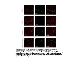
Figure e-1 A: IFN-a is not expressed in CD19+ cells, CD3+ cells, astrocytes or
by Wildboyz
oligodendrocytes. in the active areas of a chroni...
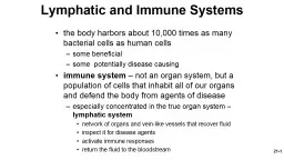
21- 1 the body harbors about 10,000 times as many bacterial cells as human cells
by alexa-scheidler
some beneficial. some potentially disease causin...
SUPPLEMENTARY FIGURES Figure S1
by patricia
Figure S2. A. B. C. D. Figure S3. A. B. C. D. Figu...
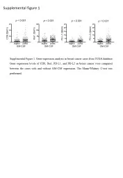
Supplemental figure 1 PD-L2 (
by eleanor
fpkm. ). PD-L1 (. fpkm. ). CD8 (. fpkm. ). Iba1 (....
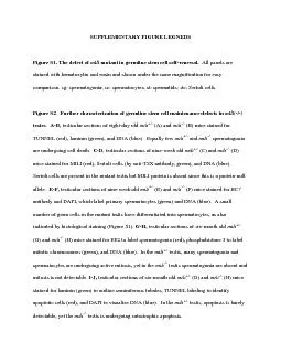
SUPPLEMENTARY FIGURE LEGNEDS Figure S1. The defect of mutant in germl
by brianna
Figure S3. MILI antibody specifically recognizes ...
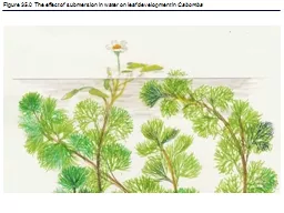
Figure 35.0 The effect of submersion in water on leaf development in
by jane-oiler
Cabomba. Figure 35.0x The effect of wind on plan...
Figure 26.0-1 Why Reproduction Matters
by phoebe-click
Figure 26.0-1a. Figure 26.0-1b. Figure 26.0-1ba. ...
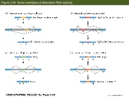
Figure 2.26 Some examples of alternative RNA splicing
by test
Figure 2.31 Degradation of casein mRNA in the pr...
Figure 35.0 The effect of submersion in water on leaf deve
by jane-oiler
Cabomba. Figure 35.0x The effect of wind on plan...
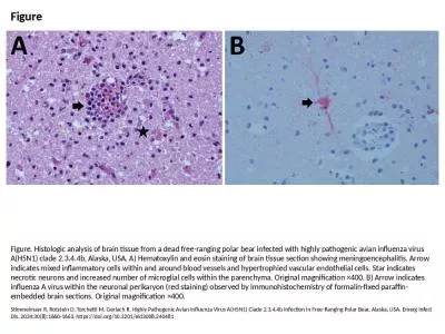
Figure Figure. Histologic analysis of brain tissue from a dead free-ranging polar bear infected wit
by august752
Stimmelmayr R, Rotstein D, Torchetti M, Gerlach R....
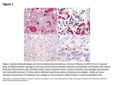
Figure 1 Figure 1. Histopathologic and immunohistochemical evidence of avian influenza A (
by landon
Keawcharoen J, Oraveerakul K, Kuiken T, Fouchier R...
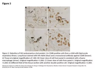
Figure 3 Figure 3. Detection of WU polyomavirus viral protein 1 in CD68-positive cells from a child
by tate
Siebrasse EA, Nguyen NL, Willby MJ, Erdman DD, Men...
Prepare for chapter three – watch this tutorial before class weds the 17th
by badra
https://sophia.smith.edu/blog/barresilab/devidetor...
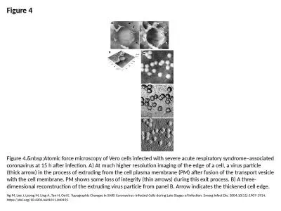
Figure 4 Figure 4. Atomic force microscopy of Vero cells infected with severe acute respir
by ella
Ng M, Lee J, Leong M, Ling A, Tan H, Ooi E. Topogr...
Chapter 14: Mind, Body and Immunity
by williams
Biology Trending, 4e. Eli Minkoff and Jennifer Hoo...
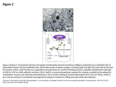
Figure 2 Figure 2. A. Transmission electron micrograph of Salmonella-induced membrane ruff
by victoria
Goosney DL, Knoechel DG, Finlay BB. Enteropathogen...
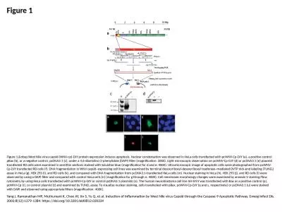
Figure 1 Figure 1. West Nile virus capsid (WNV-cp)-DJY protein expression induces apoptosi
by eleanor
Yang J, Ramanathan MP, Muthumani K, Choo AY, Jin S...
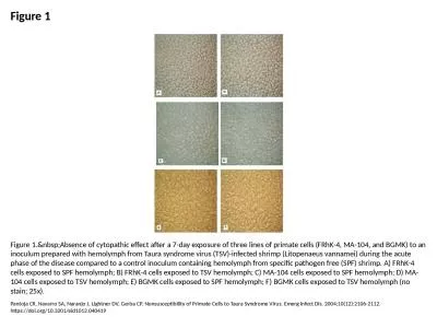
Figure 1 Figure 1. Absence of cytopathic effect after a 7-day exposure of three lines of p
by jovita
Pantoja CR, Navarro SA, Naranjo J, Lightner DV, Ge...
Figure 3 Figure 3. Scanning electron microscopy of Vero E6 cells infected with severe acut
by madeline
Ng M, Lee J, Leong M, Ling A, Tan H, Ooi E. Topogr...
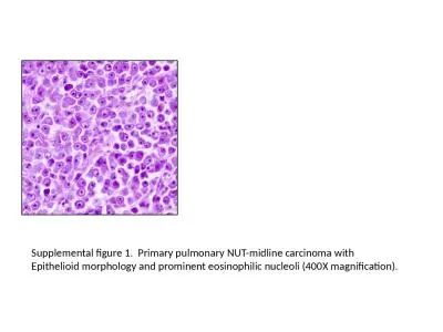
Supplemental figure 1. Primary
by jaena
. pulmonary NUT-midline carcinoma with. Epithelioi...
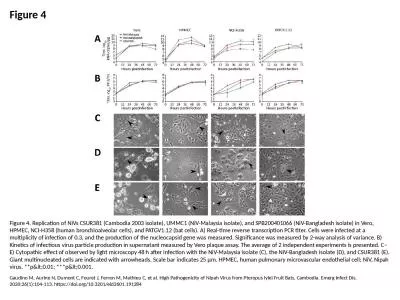
Figure 4 Figure 4. Replication of NiVs CSUR381 (Cambodia 2003 isolate), UMMC1 (NiV-Malaysia isolate
by rodriguez
Gaudino M, Aurine N, Dumont C, Fouret J, Ferren M,...
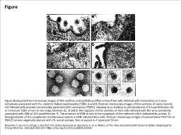
Figure Figure. Electron microscopy images of thin sections and partially purified virions
by faith
Riquelme C, Escors D, Ortego J, Sanchez CM, Uzelac...
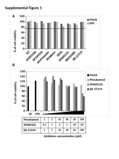
Supplemental Figure 1 Piceatannol
by williams
1. 5. 10. 30. 50. 100. SP600125. 0,1. 1. 2. 5. 10....
Drosophila melanogaster
by gabriella
Figure 8-1. The Cell Cycle ...
Iranian Journal of Pharmaceutical Sciences
by candy
2018: 14 (2): 39 - 50 www.ijps.ir Original Articl...
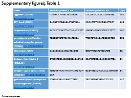
Name Forward primer 5’ – 3’
by RefreshingView
Reverse primer 5’ – 3’. (bp). Aggrecan (ACAN...
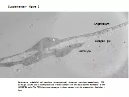
Supplementary figure 1 3d-astrocyte endothelial cell co-culture (uncompressed). Astrocytes constitu
by lucy
hCMEC. /D3 cells. The TEM shows one astrocyte in d...
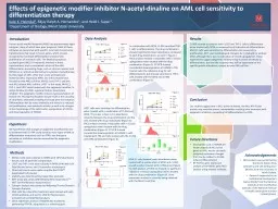
Human acute myeloid leukemia (AML) has approximately eight subtypes, many of which have poor progno
by valerie
oncoproteins. that block differentiation and prom...
Load More...